Contactless measurement of heart rate variability HRV which reflects changes of the autonomic nervous system ANS and provides crucial information on the health status of a person would. So if your heart rate is 60 beats per minute its not actually beating once every second.
 Heart Rate Variability Hrv Science For Sport
Heart rate variability The participants performed an orthostatic test 5 minutes supine followed by 5 minutes in the standing position on a regular basis between the 1 st of January 2020 and the 31 st of May 2020 22.

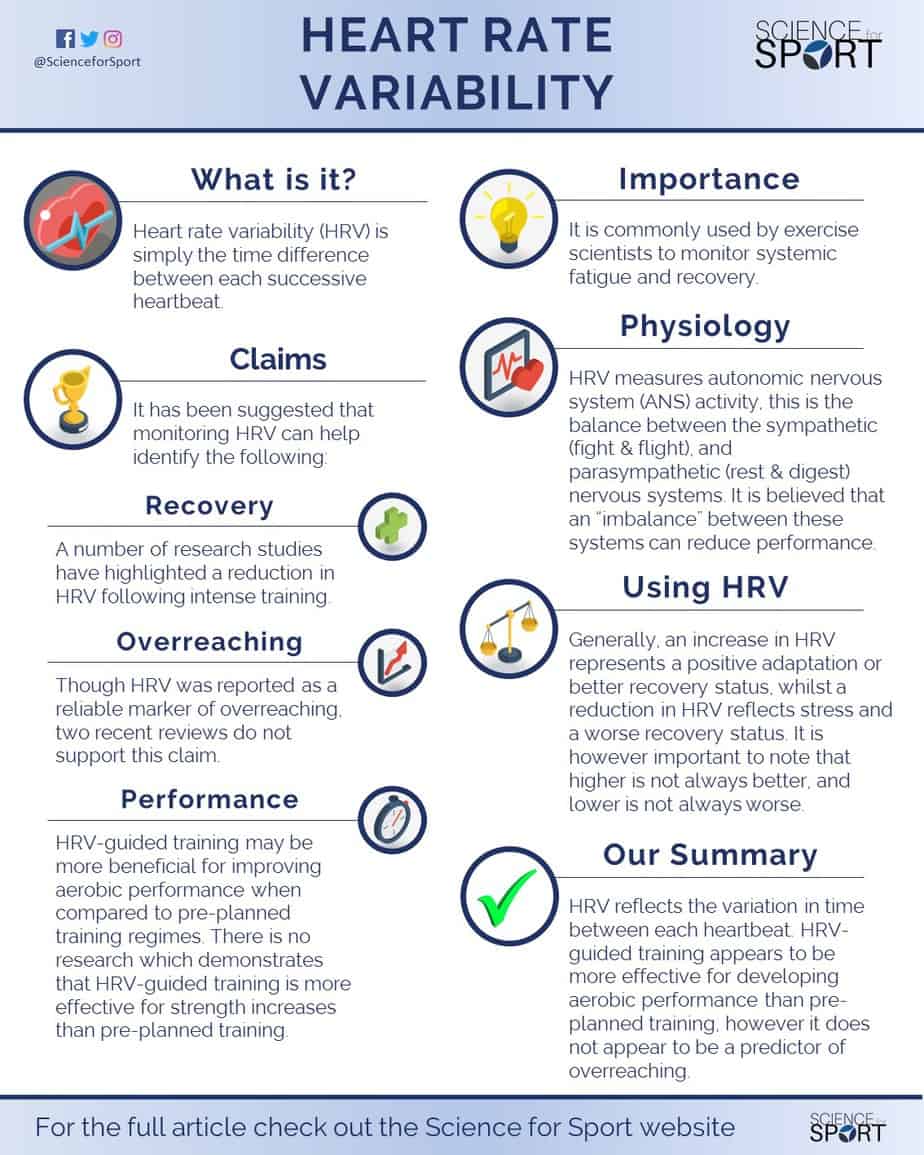
Heart rate variability test. HRV is a measure of the degree of variation in time between heartbeats. Heart Rate Variability HRV evaluates the balancing act between the sympathetic nervous system fight and flight and the parasympathetic nervous system rest and digest. If one is in a more relaxed state the variation between beats is high.
A number of published literature has reported that physiologically heart rate variability HRV in patients with postural orthostatic tachycardia syndrome POTS to be greatly confounded by age sex race physical fitness and circadian rhythm. So if the heart rate is 60 beats per minute. Within that minute there may be 09 seconds between two beats for example and 115 seconds between two others.
And since each app uses a unique scale its hard to compare the numbers across different programs. HRV is literally the difference in time between the beats of the heart. And while that may not sound that profound its actually an important metric to use if you know how to find it -- unlike heart.
Heart rate variability HRV software measures the differences between the R-peak and lengths on an electrocardiogram ECG. If a persons system is in more of a fight-or-flight mode the variation between subsequent heartbeats is low. Heart Rate Variability HRV is a 5 minute test that is non-invasive and taken while sitting comfortably in a chair.
Heart rate variability is the measurement of the autonomic nervous system ANS that is largely believed to be one of the finest objective metrics for physical strength and determine the bodys readiness to perform any action. What is Heart Rate Variability. Decreased heart rate variability and its association with increased mortality after acute myocardial infarction.
And RR is the interval between successive Rs and heart period. Heart rate variability is literally the variance in time between the beats of your heart. So good and bad HRV scores in the general population have yet to be firmly established.
HRV is an interesting and noninvasive way to identify these ANS imbalances. Heart rate variability is the amount of time between your heartbeats. What is heart rate variability.
While heart rate refers to the number of times your heart beats per minute heart rate variability HRV measures the time between each heartbeat. This orthostatic test had to be performed in the morning upon wake-up with an empty bladder and before breakfast. The simple hand electro-sensor test shows you a report on the balance between your heart and nervous system.
Heart rate variability HRV is a measure of variations in the time intervals between your heartbeats how uneven your heartbeat is. Heart rate variability in relation to prognosis after myocardial infarction. Why check heart rate variability.
Cycle length variability RR variability where R is a point corresponding to the peak of the QRS complex of the ECG wave. An imbalance in HRV is the 1 risk factor for sudden cardiac death. Heart rate variability data on healthy people is scarce.
The test lasts about 10 minutes and is painless. Crossref Medline Google Scholar. Your health care provider will place a strap around your chest that monitors heart beats.
17 Malik M Farrell T Cripps T Camm AJ. Step forward a little-known health marker called heart rate variability HRV. Heart Rate Variability or HRV is used for the calculation of Physiological Measurements such as the VO2 Max Estimator Stress Score Performance Condition Lactate Threshold and Body Battery.
Greater variation means were in a good place resilient in control of our emotions and ready for anything. The purpose of this study was to compare between POTS patients versus healthy participants in terms of heart rate HR and HRV after. First designed to track astronaut performance in the 1960s.
Other terms used include. And unlike your heart rate which you can calculate by counting your pulse heart rate variability is measured at the doctors office with an electrocardiogram ECG or EKG test that records the. Selection of optimal processing techniques.
Heart rate variability HRV is the physiological phenomenon of variation in the time interval between heartbeatsIt is measured by the variation in the beat-to-beat interval. The R-peak is the highest point and can appear like the summit of a mountain. Also known as an R-R interval this beat-to-beat interval variation is measured in milliseconds and can vary depending on a number of factors.
HRV is also used to determine sleep levels on newer Garmin devices that feature an Optical Heart Rate sensor. Or you can take a HRV test with a special machine called a heart rate variability biofeedback device. Lower variation implies we need to prioritise self-care.
With class 3 CHF your everyday activities are limited as a result of the condition. People in class 4 have severe.
Symptoms And Progression Of Heart Failure Syncardiasyncardia
The symptoms can include chest pain and shortness of breath in coronary artery atherosclerosis.

End stage heart failure symptoms. Shortness of breath that is severe and begins suddenly accompanied by coughing up a foamy pink mucus. Those symptoms experienced by patients with chronic heart failure but they begin suddenly are more severe and worsen suddenly. Even with the head elevated sleep is often interrupted by sudden spasms of breathlessness.
You may have trouble nodding off to sleep or you might wake up in the middle of the night gasping for air. Fatigue However its important to rule out any other possible causes such as anaemia not having enough red blood cells or haemoglobin problems sleeping which can be due to orthopnoea breathlessness on lying flat depression or doing more activity or exercise than theyre well enough to do. In short that means conventional heart therapies and symptom management strategies are no longer working.
Five geographically diverse tertiary care academic medical centers. In addition people in the final stages of heart failure may suffer from. Stage D heart failure is characterized by heart failure symptoms experienced when the patient is at rest even when receiving medical treatment.
Congestive Heart Failure CHF occurs when the heart is unable to pump blood fast enough resulting in swelling shortness of breath and other issues. Heart disease high blood pressure diabetes myocarditis and cardiomyopathies are just a few potential causes of congestive heart failure. Depending on the cause coughing and accompanying symptoms caused by heart failure can be experienced in different ways.
Class 2 refers to those who are largely well but need to avoid heavy workloads. The symptoms of end-stage congestive heart failure include dyspnea chronic cough or wheezing edema nausea or lack of appetite a high heart rate and confusion or impaired thinking. A retrospective analysis of data from a prospective cohort study.
Anxiety about their future. In the American Heart Association and American College of Cardiologys A-to-D staging system advanced heart failure is stage D. The main physical symptoms of heart failure at the end of life include.
The most primary symptoms of the end-stage congestive heart failure involve the inability to do any physical activity. The treatment for stage D heart failure generally involves the implantation of mechanical cardiac devices including defibrillators or pacemakers heart transplant or other types of aggressive medical therapy notes eMedicineHealth. The other symptoms associated include- Tiredness or Fatigue The heart muscles actually worn out and any activity becomes novel for the patient.
Treatments such as medications and healthier lifestyles can help people with heart failure live longer more active lives. Lying flat cannot be tolerated because the fluid-stiffened lungs cannot expand without the aid of gravity. Learn about the hospice eligibility requirements for end-stage heart failure.
Heart failure can make it hard to breathe or catch your breath when you lie in bed. A wet cough producing frothy sputum that may be tinged pink with blood 1 Heavy wheezing and labored breathing accompanied by spells of coughing A bubbling feeling in the chest or a whistling sound from the lungs. Signs and symptoms of congestive heart failure may include fatigue breathlessness palpitations angina and edema.
The abdomen ankles feet and legs fill with excess fluid in the organs and tissues leading to weight gain and difficulty performing daily tasks. Symptoms of CHF vary depending upon the organ that is affected due to lowered oxygen supply. A total of 1404 patients enrolled in the Study to Understand Prognoses and Preferences for Outcomes and.
Someone with advanced heart failure feels shortness of breath and other symptoms even at rest. Trouble navigating the health care system. To characterize the experiences of patients with congestive heart failure CHF during their last 6 months of life.
However common symptoms include. Swelling in the legs and feet caused by a buildup. What Do Symptoms of End Stage Congestive Heart Failure Look Like.
Any form of activity result in shortness of breath in the patient. Edema is a persistent and problematic symptom in the individual with congestive heart failure. The symptoms of CHF vary greatly depending on the stage and whether a person has any other medical conditions.
Treatment of End-Stage Heart Failure. This occurs as the brain receives lesser blood with each passing day. In the last stages the symptoms are quite severe and may manifest into an overwhelming feeling of weakness fatigue accompanied with constant dizziness.
Depression fear insomnia and isolation. Coughing up frothy fluid is common. Nearly 6 million Americans suffer from Congestive Heart Failure.
In end-stage heart failure no amount of activity can be undertaken without severe shortness of breath 1.
Arrhythmia which is also known as heart arrhythmia is a common disease condition that occurs when the impulses that flow through your heart and help control your heartbeat dont flow appropriately causing your heart to either beat too slow or too fast and eventually this will cause what is known as irregular heartbeats or irregular heart patterns. High blood pressure diabetes low blood sugar obesity sleep apnea and autoimmune disorders are among the conditions that may cause heart rhythm problems.
 Cancer Treatment Induced Arrhythmias Circulation Arrhythmia And Electrophysiology
An arrhythmia is a problem with the rate or rhythm of the heartbeat.

What is heart arrhythmia caused from. An irregularity in the rhythm of your heart or arrhythmia can be caused by various factors. The frequency of heart rate in one minute considered normal in an individual at rest is between 50 a to 100. High blood pressure diabetes thyroid disorders stress smoking certain medicines and consumption of alcohol or caffeine can.
Why this happens is not known. This is an arrhythmia caused by one or more rapid circuits in the atrium. Sinus node dysfunction - This usually causes a slow heart rate bradycardia with a heart rate of 50 beats per minute or less.
Arrhythmias may even be one of the more common panic attack triggers. Most anxiety-related arrhythmias have little to no effect on the heart and can occur in individuals who are extremely healthy. Things in the world.
Arrhythmias are often harmless especially when related to anxiety. WebMD explains the causes symptoms and types of arrhythmias which are changes in your heart rhythm that can be brought on by things like stress disease or certain medications. Atrial flutter is usually more organized and regular than atrial fibrillation.
An abnormal heart rhythm is when your heart beats too fast too slow or irregularly. A heart that beats irregularly too fast or too slow is experiencing an arrhythmia. Severe acute respiratory syndrome coronavirus 2 infection may cause injury to cardiac myocytes and increase arrhythmia risk.
This is also called an arrhythmia. This kills 100000 people in the UK every year. Symptoms of this type of arrhythmia include.
Heart arrhythmias uh-RITH-me-uhs may feel like a fluttering or racing heart and may be harmless. Early studies suggest that coronavirus disease 2019 COVID-19 is associated with a high incidence of cardiac arrhythmias. What Are the Causes of Arrhythmia.
Atrial fibrillation is a common cause of stroke. Having atrial fibrillation means your risk of stroke is 5 times higher than for someone whose heart rhythm is normal. As with other types of arrhythmias ventricular arrhythmias may be triggered or caused by several.
Within the heart is a complex system of valves nodes and chambers that. If other treatments are not effective a surgeon may have to remove. Cardiac arrhythmia can be benign or malignant being those.
Some common types of cardiac arrhythmias include. During an arrhythmia the heart can beat too fast too slow or with an irregular rhythm 2. Heart conditions like coronary artery disease heart attack heart valve disease and congenital heart disease may be at play.
Certain types of arrhythmia occur in people with severe heart conditions and can cause sudden cardiac death. Arrhythmia heart rate is any alteration in the rhythm of the heartstrings which can cause it to beat faster slower or just out of rhythm. This problem is most often caused by abnormalities in the hearts electrical stimulation which regulates the timing of heartbeats.
Those causes can include. There are several things that can cause arrhythmia. Also known as super ventricular tachyarrhythmia are a group of conditions which results in the heart beating faster than normal more than 100 beats per minute.
The term arrhythmia refers to any change from the normal sequence of electrical impulses causing abnormal heart rhythms 1. Scarring of heart tissue from a prior heart attack. This is why heart failure can cause swollen feet Ventricular arrhythmias causes and triggers.
The most common cause is scar tissue that develops and eventually replaces the sinus node. A palpitation is a short-lived feeling like a feeling of a heart racing or of a short-lived arrhythmia. Heart rhythm problems heart arrhythmias occur when the electrical impulses that coordinate your heartbeats dont work properly causing your heart to beat too fast too slow or irregularly.
Palpitations may be caused by emotional stress physical activity or consuming caffeine or nicotine. You will know everything you need to know about the basics about arrhythmias. But arrhythmias often make anxiety symptoms worse and may trigger panic attacks.
Unfortunately there isnt one cause to an irregular heartbeat. Atrial fibrillation is a very common irregular heart rhythm that causes the atria the upper chambers of the heart to contract abnormally. Sometimes an aneurysm or bulge in a blood vessel that leads to the heart can cause arrhythmia.
S3 results from the ventricular wall not expanding fully which causes early diastole. S1 occurs just after the beginning of systole and is predominantly due to mitral closure but may also include tricuspid closure components.
 Splitting Of The Second Heart Sound S2 Youtube
ADDITIONAL HEART SOUNDS OPENING SNAPS.

Split s2 heart sound audio. The second sound S2 is usually single. Opening snap occurs due to forceful Opening of a stenosed valve and it is described in Mitral stenosis Refer MS. Mitral Valve Prolapse.
The sound is also related to rapid filling of the ventricle. The second sound you hear is S2 and is caused by the closure of the semilunar valves SL AORTIC AND PULMONIC. S1 and the 2nd heart sound S2 a diastolic heart sound are normal components of the cardiac cycle the familiar lub-dub sounds.
Heart Sound Murmur Library. The first sound you hear is S1 and is caused by the closure of the atrioventricular valves AV TRICUSPID AND MITRAL VALVES. The pulse can be felt during systole between S1 and S2.
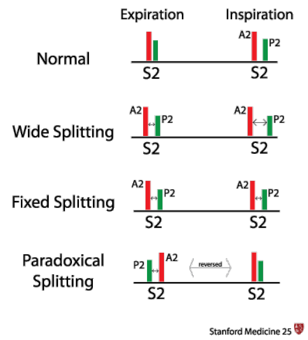
S 2 may be subdivided into aortic A 2 and pulmonary P 2 sounds as the aortic valve closes slightly before the pulmonary valve. Listen for S2 splitting. The second heart sound S2 is created by the closing of the aortic valve followed by the closing of the pulmonic valve.
Learn these sounds by selecting a topic from the table of contents below. It is caused when the closure of the aortic valve and the closure of the pulmonary valve are not synchronized during inspiration. As discussed above the second heart sound S2 is physiologically split during inspiration but not during expiration.
Basics about Heart Sounds. The splitting between A 2 and P 2 can be exaggerated by inspiration particularly in young individuals. Aortic valve closure A2 which happens first.
Careful analysis of the splitting and intensity of the second heart sound can indicate the presence of many cardiac abnormalities. Apex Area - Supine Listening with the bell of stethoscope. Its Split during expirationit is also called as Reverse splitting which.
When the aortic valve closes just before the pulmonic valve it may generate a split S2. The second sound S2 is made of two component sounds. The aortic and pulmonic valves close and cause vibrations giving rise to the second heart sound S2.
In the normal heart. ASD reversed splitting in expiration. A2 is heard widely all over the chest.
This sounds like LUB. The aortic valve closes slightly before the pulmonic and this difference is accentuated during inspiration when S2 splits into two distinct compone. The increase in intensity of this sound may indicate certain conditions.
A split S2 is a finding upon auscultation of the S2 heart sound. This type of splitting is associated with Right Bundle Branch Block a condition in which the electrical signal which causes contraction of the right ventricle is blocked. With expiration aortic valve closure A2 and pulmonic valve closure P2 may be superimposed and are rarely split as much as 004 second 4.
Heart sounds are caused by the closure of heart valves. LBBB AS coarctation PDA S3 low pitched mid-diastolic gallop rhythm. Studies have demonstrated that the aortic valve shuts first producing the first component of the second heart sound and the pulmonary valve the second component.
This heart sound is usually a normal finding in most people but can be heart in people with a right bundle branc. Next Previous screen 1 of 1 Instructions. Heart Sounds Audio Lessons Learn cardiac auscultation by taking our courses.
This may indicate impairment in the heart function. Sound is produced by the closure of the aortic and pulmonic valves cause the production of S2 sound. These courses cover abnormal heart sounds including heart murmurs third S3 and fourth S4 heart sounds and congenital conditions.
Therefore the presence of audible S2 splitting during expiration ie the ability to hear two distinct sounds during expiration is of greater significance at the bedside in identifying underlying cardiac pathology. RBBB PS VSD MR fixed splitting no respiratory variation. An S2 that remains split during both inspiration and expiration is referred to as a fixed split S2.
This is the sound of a split S1 heart sound. When theres always a delay within the closure of the pulmonic valve and theres no further delay with inspiration then fixed split S2 occurs. Usually the opening of cardiac valves does not make any sound.
Hence it is always pathological. S1 Loud MV or TV open long - shuts forcefully MS increased HR short AV conduction Soft first degree HB LBBB MR Splitting RBBB S2 Loud HT AS PHT Soft AS AR Splitting increased normal splitting wider on inspiration. The third heart sound S3 is a normal finding in children.
The S2 second heart sound is usually single. No split or a wide split indicates a problem. This example shows normal physiological splitting of the second heart sound.
Physiologically split-second heart sound. Pulmonic valve closure P2 which happens second. The second heart sound is caused by the closure of the aortic and pulmonic valves which causes vibration of the valve leaflets and the adjacent structures.
Click on the diagram of the heartsound to listen to it or stop it from playing. It is a high-pitched sound that occurs after S2. In addition to the normal components of the cardiac cycle S1 and S2 occasionally extra sounds will be present.
With persistent splitting the second heart sound is split in both inspiration and expiration although the degree of splitting is reduced in expiration. This happens normally during inspiration and in that case it is referred to as a physiologically split S2. If the S2 is split by greater than 004 second on expiration it is usually abnormal 5.
Download all sounds as mp3s.