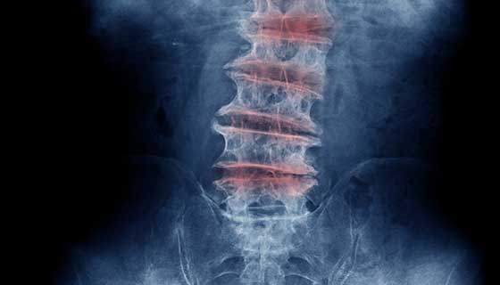
Later joint pain commonly manifests. Rheumatoid arthritis is a form of arthritis that is marked by pain or stiffness in joints including in some cases the joints of the spine.

The development of osteoarthritis in the spine is specifically referred to as spondylosis.

Arthritic changes in spine. Osteoarthritis in the spine most commonly occurs in the neck and lower back. Arthritis in the spine is one of the major sources of central spinal stenosis which is a potentially very serious condition. Wide-ranged lower back pain.
Eating a healthy diet and maintaining a healthy weight can improve symptoms and alleviate spinal. Osteoarthritis of the spine may cause stiffness or pain in the neck or back. In the normal spine the vertebrae and carilage which cushions the bones as they move are healthy and in alignment.
The most common cause of lumbar arthritis is osteoarthritis OA. Arthritis of the spine describes a host of changes in and around the joints of the spine as a result of lost disc height between the vertebrae. In the spine this breakdown affects the facet joints where the vertebrae join.
The most common type of arthritis affecting the spine is osteoarthritis also known as wear-and-tear arthritis. As with other types of osteoarthritis spondylosis is a degenerative disorder. Yet the best thing to do is to know if you have any of these spinal arthritis symptoms to get the proper medications.
It isnt a condition but rather a symptom of several forms of arthritis that affect the spine. The stiffness is worst upon waking up in the morning tends to ease with activity then worsens toward the end of the day. Lumbar spine arthritis is also known as spinal arthritis.
Presumably this is because fluid has built up in the joint due to inactivity overnight which causes more swelling. Osteoarthritis is the most common form of arthritis. Stiffness in your spine every morning.
It may also cause weakness or numbness in the legs or arms if it is severe enough to affect spinal nerves or the spinal. Osteoarthritis is the most common cause of. Cervical spondylosis is a common neck condition that arises from age-related degenerative changes in spinal joints and discs.
Degenerative changes in the spine sometimes called osteoarthritis impair the normal functioning of the spine. Osteoarthritis OA The most common form of arthritis OA occurs when the cartilage that cushions the ends of the bones where they meet to form joints breaks down or wears away. Spinal arthritis is inflammation of the facet joints in the spine or sacroiliac joints between the spine and the pelvis.
Lumbar arthritis affects the lumbar. Any time something interferes with the functioning of the spines cartilage and bone discs the spine can lose its flexibility and stiffen. Updated on November 11 2019.
Usually the low back and sometimes the neck are affected. As a result movement of the bones can cause irritation further damage and the formation of bony outgrowths called spurs. Spinal arthritis causes stiffness and low back pain.
Although most cases of arthritic debris in the central canal do not become symptomatic advanced cases or cases where the canal diameter has already been compromised by injury or a congenital condition may elicit truly catastrophic symptoms for the patient. Symptoms of spondylosis tend to come on gradually as your spine changes. Most people over age 60 have degenerative changes in their spine consistent with osteoarthritis but for perhaps 85 of them there is no pain or stiffness.
You may know cervical spondylosis as neck osteoarthritis or degenerative disc disease of the neck. With aging comes wear and tear on the bodys joints including those of the spine. The phrase degenerative changes in the spine refers to osteoarthritis of the spine.
Back pain which occurs all of a sudden then fades away. By Jennifer Cuthbertson Osteoarthritis OA is the most common form of arthritis that affects the back. Although osteoarthritis usually occurs during old age specific conditions such as osteoporosis spondylitis osteomyelitis or a tumor could also be at the root of spinal pain.
Doctors may also refer to it as degenerative arthritis or degenerative joint disease. It can occur anywhere along the spine. The spine is divided into five sections the cervical thoracic lumbar sacrum and coccyx.
Conversely an X-ray conducted during the early stages of osteoarthritis may not yet show any visible damage to the joints but the patient may have symptoms. Theres a special name for spinal osteoarthritis. It may be related to wear and tear autoimmune disorders infection and other conditions.
Lumbar spine arthritis is typically seen alongside other spinal disorders. Making healthy lifestyle changes can make it easier to live with osteoarthritis of the spine. Its present in some degree in most people after 40 years of age.
Sometimes the inflammation may also affect the sites where ligaments and tendons attach to the bones of the spine. Early symptoms of rheumatoid arthritis include excessive fatigue fever reduced appetite and weakness. Pain numbness and swelling in several parts of your body such as neck knees hips heels or shoulders.
Hyperkalemia is not always expressed with EKG changes. Hyperkalaemia causes a rapid reduction in resting membrane potential leading to increased cardiac depolarization and muscle excitability which in turn causes EG changes 1.
ECG changes have a sequential progression which roughly correlate with the potassium level but with the caveats mentioned above 2 Early ECG changes of hyperkalemia typically seen at a serum.

Ecg changes in hyperkalemia. In a prospective series 46 of patients with hyperkalemia were noted to have electrocardiographic changes but no clear criteria were presented. Recognition of the ECGEKG changes of hyperkalemia can save lives. In most cases EKG changes have good correlation with the degree of hyperkalemia.
But the levels at which ECG changes are seen are quite variable from person to person. Any acute or chronic kidney disease or dysfunction interrupts the delicate homeostasis and causes hyperkalemia. Kayexalate takes time to work.
With severe hyperkalemia effect on RMP becomes prominent. IV insulin f ollowed by glucose will shift potassium intracellularly and is an effective treatment for severe hyperkalemia. Minimal potassium level 55 to 65 mEqL.
Design setting participants measurements. In patients with mild hyperkalemia potassium conductance IKr through potassium channels is increased which tend to shorten the AP duration and on the ECG causing tall tented T waves. Pt with ESRD getting dialysis 3 times per week presents with K 9 on.
The effect is transient but the calcium injection can be repeated until measures to correct the hyperkalemia are undertaken. It produces predictable changes on the ECGEKG. There is a predictable EKG progression as the serum potassium becomes more elevated.
Electrocardiogram Findings in Hyperkalemia. Hyperkalemia serum K 55 mmoll is a life-threatening medical emergency. Hyperkalemia causes a rapid reduction in resting membrane potential leading to increased cardiac depolarization and muscle excitability.
Classic teaching of the chronological ECG changes of hyperkalemia include. ECG findings in hyperkalemia Hyperkalemia can result in a variety of presentationsincluding asymptomatic dyspnea nauseavomiting diarrhea weakness chest pain missed dialysis or cardiac arrest. Prolongation of PR interval.
12 Hyperkalemia is defined as a serum level of potassium greater than 55 mEqL. Potassium of 7 is severe hyperkalemia. Where did the evidence for order of ECG changes of hyperkalemia come from.
Charts were reviewed for patients who were admitted to a community-based hospital with a diagnosis of hyperkalemia. Hyperkalemia serum K 55 mmoll is a life-threatening medical emergency. Most patients have risk factors including CKD CHF DM or medications like ACE inhibitors or potassium-sparing diuretics.
It produces predictable changes on the ECGEKG. Recognition of the ECGEKG changes of hyperkalemia can save lives. Tall peaked tented T waves T wave larger than R wave in more than 1 lead.
Loss of P wave Sine Wave Asystole. The electrocardiographic ECG or EKG changes with hyperkalemia do not consistently follow a stepwise dose-dependent pattern. There are five ECGEKG changesgroups of changes associated with hyperkalemia which you must be able to recognise.
Peaked T waves usually the earliest sign of hyperkalaemia. Although the electrocardiogram ECG is not considered a reliable indicator of mild to moderate hyperkalemia profound elevations of serum potassium concentration generally produce classic ECG manifestations. You want to urgently bring down the potassium.
There are five ECGEKG changesgroups of changes associated with hyperkalemia which you must be able to rec. ECG changes are usually progressive and may include. Hyperkalemia with ECG changes.
It produces predictable changes on the ECGEKG. Electrophysiologic basis of ECG changes. It can be further broken down as follows.
Associated with minor ECG changes. We report two cases of severe hyperkalemia 90 mEqL in which the ECGs did not reveal the expected manifestations of hyperkalemia. Hyperkalemia serum K 55 mmoll is a life-threatening medical emergency.
As the severity of hyperkalemia increases the QRS complex widens and the merging together of the widened QRS complex with the T wave produces the sine wave pattern of severe hyperkalemia. Tall tented T waves. In this one critical minute we talk about the ECG changes in hyperkalemiaThe first changes that occur as potassium levels rise are the peaking of T-waves.
This in turn can cause electrocardiographic ECG or EKG changes 10. In the presence of ECG changes and hyperkalemia intravenous calcium usually as gluconate since chloride is chemically more toxic if extravasated results prompt reversal of ECG changes. Effects of hyperkalaemia on the ECG Serum potassium 55 mEqL is associated with repolarization abnormalities.
EKG changes also depend on the rate of increase in potassium concentration. Recognition of the ECGEKG changes of hyperkalemia can save lives.
