What New Coronavirus Looks Like Under The Microscope The images were made using scanning and transmission electron microscopes at the National Institute of Allergy and Infectious Diseases. Backscattered electron detector BSED.

It is a huge magnification of the common dust of industrial outdoor environments.

Scanning electron microscope images. Electromagnetic lenses Condenser lens Objective lens. The high-resolution three-dimensional images produced by SEMs provide topographical morphological and compositional information makes them invaluable in a variety of science and industry applications. The electron beam is scanned in a raster scan pattern and the position of.
Electronic Gun For the source of the electron. What youre seeing above is a scanning electron microscope image in false colour showing the COVID-19 virus from a patient in the US. As the intensity of the generated secondary electrons varies depending on the angle of the incident electrons onto the specimen surface subtle variations in the roughness of the surface can be expressed according to the signal intensity.
The electron source and electromagnetic lenses that generate and focus the beam are similar to those described for the. See more ideas about microscopic photography scanning electron microscope electron microscope. Feb 1 2017 - Explore Sara Kuntzs board Scanning Electron Microscope Images on Pinterest.
The scanning electron microscope SEM uses a focused beam of high-energy electrons to generate a variety of signals at the surface of solid specimens. - scanning electron microscope stock pictures royalty-free photos images. Colorful image for tops t-shirts skirts scarves leggings socks.
Thousands of new high-quality pictures added every day. Component or instrument used in scanning electron microscope. See more ideas about scanning electron microscope electron microscope images electron microscope.
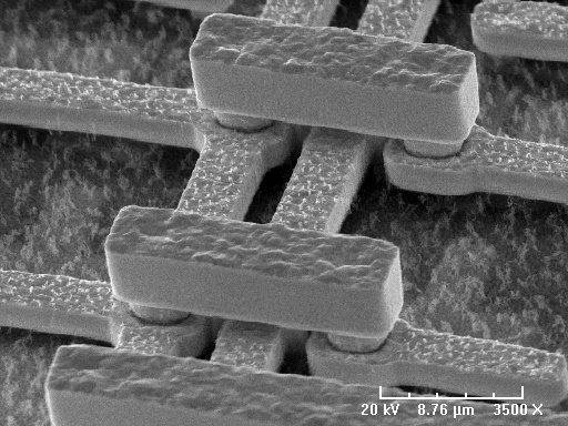
Common industrial dust Technically this image is not a photograph since it was not originated by light photo but by an electron beam. Scanning electron microscopy SEM is a technique that provides high-resolution electronic images of the surface of different materials by scanning with an electron beam. The electrons in the beam interact with the sample producing various signals that can be used to obtain information about the surface topography and composition.
X-rays or light detector. A Scanning Electron Microscope SEM is a powerful magnification tool that utilizes focused beams of electrons to obtain information. Colourised sem image of atropa pollen showing the hard coat which protects the sperm cells during the movement from stamens of producing flower to the pistil of the receiving flower.
The signals that derive from electron-sample interactions reveal information about the sample including external morphology texture chemical composition and crystalline structure and orientation of materials making up the sample. Apr 22 2017 - Scanning Electron Microscope Images - Unbelievable how life really is up close. The Scanning Electron Microscope SEM normally detects secondary electrons to form an image for observation.
The viral particles are coloured yellow as it emerges from the surface of a cell which is coloured blue and pink. A scanning electron microscope SEM scans a focused electron beam over a surface to create an image. NIAID-RML The image above was captured with a transmission electron microscope.
25 Amazing Electron Microscope Images Writen by Bogdan Comments Off on 25 Amazing Electron Microscope Images All the common objects are kinda boring when you look at them but the situation changes when an awesome Electron Microscope comes in the scene. A scanning electron microscope SEM is a type of electron microscope that produces images of a sample by scanning the surface with a focused beam of electronsThe electrons interact with atoms in the sample producing various signals that contain information about the surface topography and composition of the sample. Secondary electron detector SED.
The image is modified and credit goes to Wikimedia. Atropa is a genus of the nightshade family. The image was captured by an Hitachi ultra-high-resolution Analytical FE Scanning Electron Microscope SU-70.
This scanning electron microscope image shows SARS-CoV-2 yellow the virus that causes COVID-19 emerging from the surface of cells bluepink cultured in the lab. Find scanning electron microscope stock images in HD and millions of other royalty-free stock photos illustrations and vectors in the Shutterstock collection. Scanning electron microscope SEM type of electron microscope designed for directly studying the surfaces of solid objects that utilizes a beam of focused electrons of relatively low energy as an electron probe that is scanned in a regular manner over the specimen.
In addition the contrast of the images provides information about the composition of the surface sample as its different elements emit different amounts of characteristic electrons. Scanning electron microscopy SEM in particular has given us some striking images over the years to tantalize our visual senses.