Schematic overview of single-cell ATAC-seq assays and analysis steps. In this tutorial we will show how to use scHINT an extended version of HINT-ATAC to compare transcription factor binding activity based on differential footprinting.
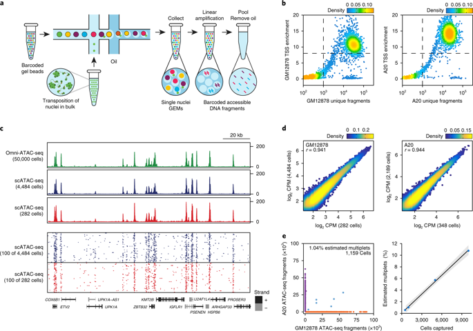 Massively Parallel Single Cell Chromatin Landscapes Of Human Immune Cell Development And Intratumoral T Cell Exhaustion Nature Biotechnology X Mol
Assay for single-cell transcriptome and accessibility regions ASTAR-seq which coincidently was developed at ASTAR in Singapore uses an integrated micro fluidic chip Fluidigm C1 to compartmentalize the cells similar to the first scATAC-seq Xing et al.

Single cell atac seq. Online ahead of print. Single-cell ATAC-seq scATAC-seq profiles the chromatin accessibility landscape at single cell level thus revealing cell-to-cell variability in gene regulation. In this article we focus on the single-cell ATAC-Seq scATAC-Seq technology to describe how it works and highlight the benefits and the drawbacks of scATAC-Seq relative to bulk ATAC-Seq.
To study such changes at the single-cell level we analyzed single-cell RNA-seq scRNA-seq and matched scATAC-seq data from primary remission andor relapse samples obtained from three pediatric AML patients enrolled in the AAML1031 clinical trial Alpenc et al. Integrative Single-Cell RNA-Seq and ATAC-Seq Analysis of Human Developmental Hematopoiesis. Cells are then tagmented with Tn5 within the chip then the RNA is reverse transcribed and amplified with biotinylated primers.
In addition the Cicero package provides an extension toolkit for analyzing single-cell ATAC-seq experiments using the framework provided by the single-cell RNA-seq analysis software Monocle. This vignette provides an overview of a single-cell ATAC-Seq analysis workflow with Cicero. Single-cell ATAC-seq scATAC-seq profiles the chromatin accessibility landscape at single cell level thus revealing cell-to-cell variability in gene regulation.
Single cells from 15 fetuses were processed for scRNA-seq using the SmartSeq2 protocol Picelli et al 2014 Figure 1AOverall 4504 cells passed quality control QC with an average of 3600 genes per cell and 670000 reads per cell Figures S2AS2C S2K and S2LTo exclude technical batch effects we merged the datasets from all samples and tissues using autoencoders AEs and. However the high dimensionality. The ATAC-Seq method is a genome-wide NGS-based assay that characterizes chromatin states in cell and tissue samples.
Single cell ATAC-Seq reveals cell type-specific transcriptional regulation and unique chromatin accessibility in human spermatogenesis. An alternative technique that does not require single cell isolation is combinatorial cellular indexing. Single-cell ATAC-seq data for human hematopoietic cell types were obtained from GEO GSE96769.
Here we report scFAN single-cell factor analysis network a deep learning model that predicts genome-wide TF binding profiles in individual cells. Analysis of single-cell chromatin accessibility data allows us to detect novel cell clusters while how to interpret these clusters remains a challenge. Next Article Decoding Human Megakaryocyte Development.
Single-cell ATAC-seq data for mouse brain and thymus were obtained from GEO GSE111586. This protocol for performing single-cell ATAC-seq was developed using the ICELL8 Single-Cell System by the Greenleaf lab at Stanford University in consultation with Takara Bio USA Inc. Here the authors characterize mouse CPCs marked by Nkx25 and Isl1 from E75 to E95 by single cell RNA-seq and ATAC-seq showing fate transitions involve distinct open chromatin state.
A Please direct any requests for more information to the authors. ScFAN is pretrained on genome-wide bulk assay. 1 Single cells are individually barcoded by a split-and-pool approach where unique barcodes added at each step can be used to identify reads originating from each cell 2 microfluidic droplet-based technologies provided by 10X Genomics and BioRad are used to extract and.
Modifications to the ATAC-seq protocol have been made to accommodate single-cell analysis. Single-cell ATAC-seq data preprocessing. Comparisons between bulk ATAC-seq and single cell ATAC-seq from cell lines indicates that single cell ATAC-seq recovers 90 of peaks recovered by bulk ATAC-seq.
Using the 10X Genomics single-cell platforms we profiled a. Single-cell ATAC-seq data for GM12878 and K562 cells were obtained from GEO GSE65360. Microfluidics can be used to separate single nuclei and perform ATAC-seq reactions individually.
However the high dimensionality and sparsity of scATAC-seq data often complicate the analysis. Single cell ATAC-seq also provides the resolution to look at chromatin profiles at a cell-type specific level. ATAC-Seq is an assay for interrogating the entire genome for accessibility to DNA binding proteins in a single experiment.
While traditional bulk assay for transposase-accessible chromatin sequencing ATAC-Seq offers valuable insights into genome-wide epigenetic regulation cells in heterogeneous populations are combined into a single transposition reaction obscuring key chromatin signatures. A Single-cell ATAC libraries are created from single cells that have been exposed to the Tn5 transposase using one of the following three protocols. In particular ATAC-Seq is used to identify regions of the genome that have open chromatin states that are generally associated with sites undergoing active transcription.
Single cell ATAC-Seq reveals cell type-specific transcriptional regulation and unique chromatin accessibility in human spermatogenesis Xiaolong Wu Xiaolong Wu Medical School Institute of Reproductive Medicine Nantong University Nantong 226001 Jiangsu China. Previous Article Dcaf11 activates Zscan4-mediated alternative telomere lengthening in early embryos and embryonic stem cells. With this approach single cells are captured by either a microfluidic device or a liquid deposition system before tagmentation.
In collaboration with Jay Shendures lab and scientists at Illumina we recently developed sci-ATAC-seq a single-cell ATAC-seq protocolOur initial study led by Darren Cusanovich explored variation in chromatin accessibility both between and within populations of.
A complete blood count CBC also known as a full blood count FBC is a set of medical laboratory tests that provide information about the cells in a persons bloodThe CBC indicates the counts of white blood cells red blood cells and platelets the concentration of hemoglobin and the hematocrit the volume percentage of red blood cells. 4254 million cells per µL of blood for females.
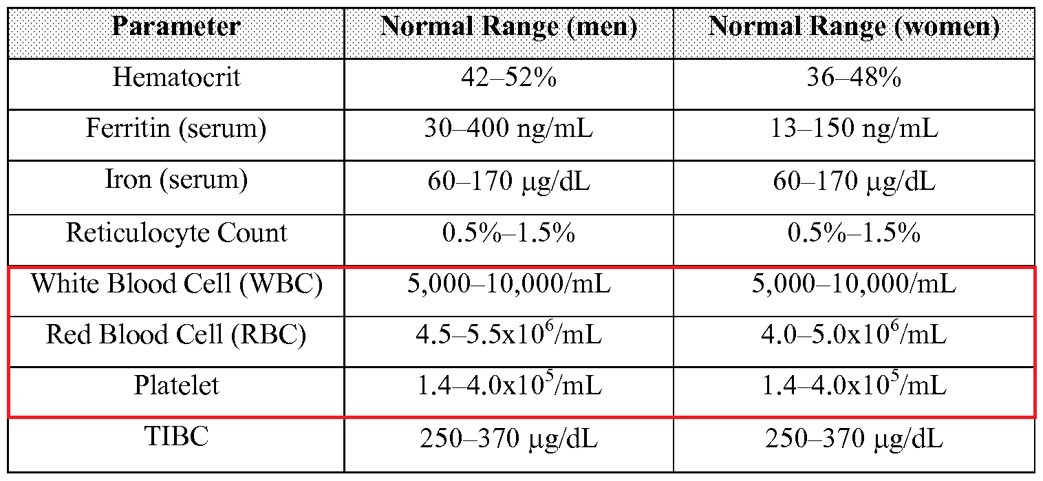 Which Type Of Blood Cell Is The Most Abundant In The Human Body Lymphocytes Basophils Erythrocytes Neutrophils Or Platelets Socratic
In children the threshold for high red blood cell count varies with age and sex.
Red blood cell count. A Quick Science Question. Red blood cells carry oxygen in the blood. Eating foods rich in 5 ingredients may help to increase your red blood cell count including iron folic acid vitamin B12 copper and Vitamin A.
For women 42 to 54 million red blood cells per microliter of blood. High red blood cell count may be caused by low oxygen levels kidney disease or other problems. 4761 million cells per microliter µL of blood for males.
The test can help diagnose different kinds of anemia low number of RBCs and other conditions affecting red blood cells. The red blood cell RBC count is used to measure the number of oxygen-carrying blood cells in a volume of blood. Inflammation and nutrient deficiencies can reduce red blood cell numbers or their ability to effectively deliver oxygen.
When a person has low red blood cells. What is a red blood cell count. What Causes Low Red Blood Cell Count.
A red blood cell RBC count is typically ordered as part of a complete blood count CBC and may be used as part of a health checkup to screen for a variety of conditions. Doctors typically find a high red blood cell count during tests for another health issue. The red blood cell indices which indicate the.
Knowing why your doctor bought an RBC count and what the results of your RBC count mean is important in understanding how it associates with your health. Your body may increase red blood cell production to compensate for any condition that results in low oxygen levels including. These ranges can vary depending on the testing lab.
The red blood cell count or RBC count lets you know if you have a low amount of red blood cells which is known as anemia or a high amount which is known as polycythemia. Disease that damages kidney blood vessels Alport syndrome. It is one of the key measures we use to determine how much oxygen is being transported to cells of the body.
Its also known as an erythrocyte count. Heart disease such as congenital heart disease in adults Heart failure. The most common cause of a low red blood cell count is the deficiency of iron in the body and the condition is termed as anemia.
Normal red blood cell counts are. Which Important Mineral Does the Haemoglobin Present in RBC Contain. This condition is called anemia.
This number however may vary depending upon the age of the individual and the testing laboratorys recommendations. This test may also be used to help diagnose andor monitor a number of diseases that affect the production or lifespan of red blood cells. For men 47 to 61 million red blood cells per microliter of blood.
Discovering anemia is often the starting point to diagnosing an underlying condition. Maintaining a normal red blood cell count is essential. A normal range in adults is generally considered to be 435 to 565 million red blood cells per microliter mcL of blood for men and 392 to 513 million red blood cells per mcL of blood for women.
Other conditions that may require an RBC count are. There are many possible causes of low red blood cell count such as chronic blood loss leading to iron deficiency anemia acute blood loss or hereditary disorders. A lack of iron in the diet and perhaps other minerals and nutrients is the most common cause of a low red blood cell count.
The RBC count is almost always part of a complete blood count test. The primary function of the red blood cell is to transport oxygen to all parts of the body. A low red blood cell count means you have anemia a condition that could be caused by a variety of factors like blood loss genetic disorders cancer treatments and other causes.
For children 40 to 55 million red blood cells per microliter of blood. Normal red blood cell count for an adult male is 47-61 Mul and for an adult female it is 42-54 Mul. However having a high red blood cell count can also have a similar oxygen-depleting effect.
A typical human red blood cell has a disk diameter of approximately 6282 µm and a thickness at the thickest point of 225 µm and a minimum thickness in the centre of 081 µm being much smaller than most other human cellsThese cells have an average volume of about 90 fL with a surface area of about 136 mm 2 and can swell up to a sphere shape containing 150 fL without membrane. Anemia can be cured by increasing the intake of iron-rich foods in your diet. A red blood cell count is a blood test that your doctor uses to find out how many red blood cells RBCs you have.
Normal RBC counts range from 47 to 61 million cells per microliter mcL for men and 42 to 54 million cells per mcL for women. A red cell RBC count is a type of blood test that can supply details about how many red blood cells are in an individuals blood. 4055 million cells per µL of blood for children.
T Cell Functional Assays. T cell exhaustion is a phenotypic and functional state of a T cell.
 Functional Profile Of Cd8 And Cd4 T Cells Response To Zikv Infection Download Scientific Diagram
Unlike the conventional T cells described in Basic Protocol 1 CD4CD25.

T cell functional assay. The supernatant can simultaneously be collected from the T cell proliferation cultures to assess cytokine production using Cytokine Bead Arrays or AlphaLISAs in our cytokine production assays. The conventional PBMC assays require large volume of blood samples when compared to WB assays. CD4CD25 regulatory T cells Treg cells prevent T cell-mediated autoimmune diseases in rodents.
To develop a functional Treg assay for human blood cells we used FACS- or bead-sorted CD4CD25 T cells from healthy donors to inhibit anti-CD3CD28 activation of CD4CD25- indicator T cells. T cell functional assays. In vitro functional assay for the study of additivesynergistic effects of compounds of interest for adjunctive or combination therapy.
Upregulation of activation markers on the cell surface 3. Naive T cells or antigen-specific memory T cell pools are often present at low frequency in peripheral blood which makes these populations hard to study in terms of phenotype and functionality directly ex vivo. Measures the frequency of antigen-specific T cells secreting numerous cytokineschemokines eg.
In these assays the lysis of target cells by T cells can be measured. Despite being invented half a century ago Timonen and Saksela 1977 the 51 Cr release assay is still being used to measure T cell-mediated cytotoxicity because of its high sensitivity and low cell number requirements. Human peripheral blood mononuclear cells PBMCs are isolated from whole blood.
Assay readouts include cytokine measurement by Flow Cytometry or ELISA. Particularly our experiments can run for several days which makes this cytotoxicity assay much more sensitive than traditional methods such as the chromium release assay. Each mature T cell will ultimately contain a unique TCR that reacts to a random pattern allowing the immune system to recognize many different types of pathogens.
This addition will also describe the assay in which CD4CD25T cells are cocultured with conventional T cells in order to assess their suppressive function. LakePharma offers a variety of assays for characterizing T-cell activity with candidate therapeutics. Induction of cytotoxicity or cytokine secretion 5.
Two different assay methods exist to analyze the T-lymphocyte functions namely Peripheral Blood Mononuclear Cell PBMC assays and Whole Blood WB assays. Clonal expansion of T cells 2. T cell activation requires at least two signals to become fully activated.
The conditions required to induce proliferation are described. T cell assays can be utilized to identify and measure a recall or memory response in PBMCs derived from subjects who have been exposed to a given biologic product either as therapy or within the within the context of a clinical trial. Our services include long-term cytotoxicity assays to reveal T cell killing in cell cultures and drug-mediated cytotoxic effects.
Furthermore we can make use of a broad range of cell lines derived from both cancer and healthy patients. The TCR consists of two major components the alpha and beta chains. Is more sensitive than intracellular cytokine staining ICS.
The first occurs after engagement of the T cell antigen-specific receptor TCR by the antigen-major histocompatibility complex MHC and the second by subsequent engagement of co-stimulatory molecules. Finally our third most commonly performed functional assay is the T cell cytotoxicity assay. T-cell activation and proliferation assays include stimulation with staphylococcal enterotoxin B SEB cytomegalovirus CMV recall antigen and anti-CD3 antibodies.
Induction of apoptosis One of the most common ways to assess T cell activation is to measure T cell proliferation upon in vitro. LakePharma offers a variety of assays for characterizing T-cell activity with candidate antibodies. This protocol is designed and describes methods to overcome these limitations by allowing for the detection of antigen-specific killing of a target cell by CD8 T cells in vivo.
It is characterised by increased cell surface expression of checkpoint inhibitors and a reduced functional capacity. Differentiation into effector cells 4. This is accomplished by merging a vaccination model with a traditional CFSE-labeled target killing assay.
Cells do not proliferate to TCR stimuli alone. Exhausted T cells exhibit reduced proliferation a reduction in cytokine production and function. Measures the frequency of IFN-g-secreting T cells in peripheral blood mononuclear cells PBMC.
Finally this addition will describe the culture conditions for the activation and expansion of CD4CD25 cells. Features of Our Services. Sorting of cells using pentamerpeptide strategies and subsequent expansion using in vitro T cell assays allows us to assess a therapys ability to modulate the function and.
A critical step in T cell maturation is making a functional T cell receptor TCR. T Cell Activation Functional Assay. This addition will also describe the assay in which CD4CD25T cells are co-cultured with conventional T cells in order to assess their suppressive function.
For the T cell proliferation assay propagation of T cells is measured by CFSE dilution or Cell Titer Glo following the stimulation period. The conditions required to induce proliferation are described. Primary T-Cell Functional Assays Primary T-Cell Functional Assays T-cell based immunotherapy strategies have become increasingly important in disease treatments that harness the human immune system.
DM can be difficult to diagnose not only clinically but a. Doctor gives it approx.
 Desmoplastic Melanoma Atypical Spindle Cells In A Dense Fibous Matrix Download Scientific Diagram
Desmoplastic melanoma is a variant of spindle cell melanoma where there are varying proportions of spindle cells and desmoplastic cells present in the histology.
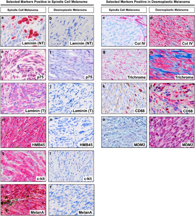
Desmoplastic spindle cell melanoma. Desmoplastic melanoma DM and spindle cell melanoma SCM are rare morphologic variants of melanoma. Spindle cell melanoma SCM is a rare morpholog - ical subtype of melanoma which is relatively uncharacterized. Desmoplastic malignant melanoma A rare variant of spindle cell melanoma John Conlev MD Departments of Pathology Division of Surgical Pathology and the Head and Neck Service ColumbiaPresbyterian Medical Center and the College of Physicians and Surgeons of Columbia University New York N Y.
Larger in size 4mm deeper Breslow IV spindle-cell makeup more frequent in older males usually found on head and neck lower incidence of lymphatic involvement despite their size Dads SLNB was negative which of course could be a cooincidence. Spindle cell melanoma SCM is a rare subtype of malignant melanoma composed of spindled neoplastic cells arranged in sheets and fascicles The diagnosis of SCM is challenging as SCM may occur anywhere on the body and frequently mimics amelanotic lesions including scarring and inflammation 24Histologically cytologic features of SCM are indistinct and often confused. Desmoplastic and spindle-cell malignant melanoma.
Adv Anat Pathol 22227-241. Spindle cell melanoma and desmoplastic melanoma differ clinically in prognosis and therapeutic implications. The clinical histologic and immunohistologic features of 22 desmoplastic melanomas DMM 10 mixed desmoplastic and spindle-cell melanomas DMMSMM and two cellular spindle-cell melanomas SMM were studied.
When reading the characteristics of desmoplastic melanoma my Dads tumor pretty much fits to a T. DM was first described as a variant of SCM in 1971 defining as a melanoma composed of spindle cells and abundant collagen. There is a difference in recurrence rates lymph node metastases and mortality.
However because of partially overlapping histopathological features diagnostic distinction of spindle cell from desmoplastic melanoma is not always straightforward. Factor XIIIa may be positive in desmoplastic melanoma 11. First described in 1971 Cancer 197128914.
Hello My husband had a lump in his head since May 2011. Desmoplastic melanoma often involves nerve fibres when it is called neurotropic melanoma. Conley J Lattes R Orr W 1971 Desmoplastic malignant melanoma a rare variant of spindle cell melanoma.
1 however the term spindle cell melanoma can either serve as an umbrella term for the spindle cell and. Frydenlund N et al2015 Desmoplastic melanoma neurotropism and neurotrophin receptors--what we know and what we do not. Desmoplastic melanoma is currently defined as a subtype of spindle cell melanoma.
Further studies are needed to clarify the clinical manifestations risk factors treatment management and prognosis of spindle cell melanoma. The clinical histologic and immunohistologic features of 22 desmoplastic melanomas DMM 10 mixed desmoplastic and spindle-cell melanomas DMMSMM and two cellular spindle-cell melanomas SMM were studied. Desmoplastic melanoma DM is a variant of spindle cell melanoma typically found on chronically sun-damaged skin of older individuals.
The malignant cells within the dermis are surrounded by fibrous tissue. Has been seen in desmoplastic melanoma. The aim of the present study was to investigate the incidence of SCM its general demographics basic clinico-pathologic features treatment outcomes and diseasespecific prognostic factors.
These represent a spectrum of subtypes that have variable spindle cell cellularity and collagen density. Rare variant of spindle cell melanoma seen in older adults in sun exposed skin. The dermatologist checked the lump in November 2011 and thought.
Spindle cell desmoplastic melanoma Often have overlying melanoma in situ 70 of cases usually lentigo maligna type May have clustered or theque-like areas Diffusely and strongly positive for S100 and SOX10 MPNST often shows only weakfocal staining Usually negative for HMB-45 tyrosinase and MART-1Melan-A Epithelioid melanoma. Early diagnosis can be challenging because it is often amelanotic and has a predominantly dermal component. Very rare in conventional melanomas and non-desmoplastic spindle cell melanomas but is commonly seen in desmoplastic melanoma 11.
Early diagnosis can be challenging because it is often amelanotic and has a predominantly dermal component. Desmoplastic Spindle Cell- need some advise. He had a surgery in February 2012 and we just got a result that the type of cancer is spindle Cell Melanoma.
A type of nodular vertical growth melanoma with scanty spindle cells prominent desmoplastic stroma and often minimal atypia. 50 50 chance this has spread already. SCM cases were sampled from the.
Patients ranged in age from 35 to 91 years mean 67 and included 23 men and 11 women. Desmoplastic melanoma DM is a variant of spindle cell melanoma typically found on chronically sun-damaged skin of older individuals. Desmoplastic melanoma is a rare form of invasive melanoma a skin cancer that arises from pigment cells melanocytes.
Desmoplastic melanoma and spindle cell melanoma are two distinct melanoma subtypes that differ clinically and histologically.
Giant cell myocarditis is treated with a combination of systemic corticosteroids and another immunosuppressive agent such as cyclosporine muromonab-CD3 or azathioprine. The United Kingdom is a constitutional monarchy comprising much of the British Isles.
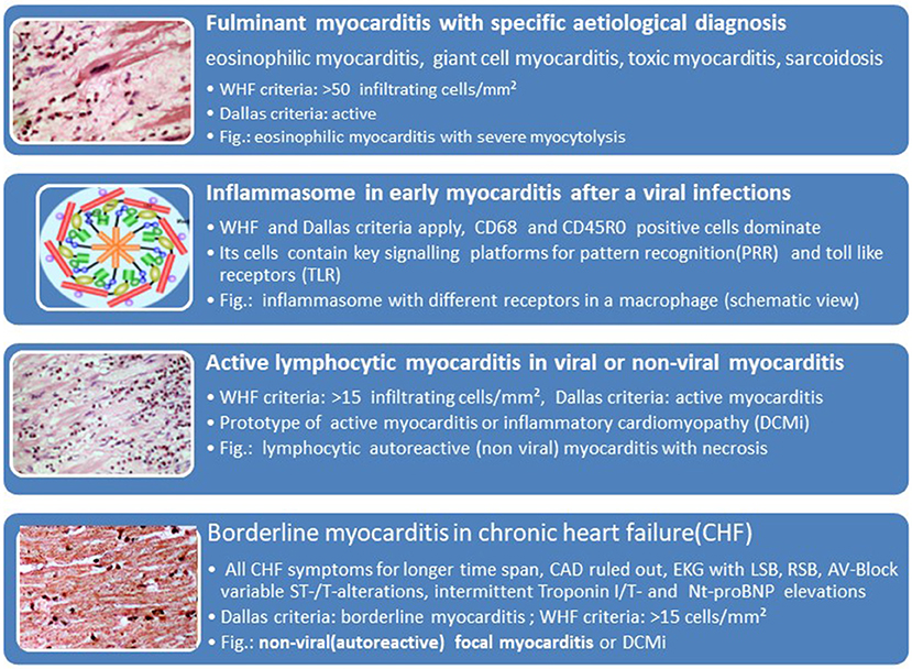 Frontiers Cardio Immunology Of Myocarditis Focus On Immune Mechanisms And Treatment Options Cardiovascular Medicine
This case highlights emerging concepts in the management of giant cell myocarditis including subacute presentations challenges in diagnosis and treatment modalities in the modern era.
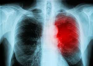
Giant cell myocarditis treatment. Current therapy rests on multiple-drug immunosuppression but its prognostic influence remains poorly known. 1860 June 26 1997 The New England Journal of Medicine IDIOPATHIC GIANT-CELL MYOCARDITIS NATURAL HISTORY AND TREATMENT L ESLIE T. Giant Cell Myocarditis Treatment is a medical procedure surgery that requires coordination between specialist surgeons anesthetists and various other specialist medical professionalsThis type of Cardiology procedure treatment can be very expensive given the extent of everything involved for example the skill.
1 In many cases clinical course is acute to fulminant and time span between clinical presentation and start of treatment should be as short as possible. In general giant cell myocarditis is a rapidly fatal disorder that may respond to certain immunosuppressive drugs or heart transplantation. The disorder most often occurs in young adults.
The following case describes the management of a patient with an unusual presentation of giant cell myocarditis over a 10 year course of advanced heart failure therapies and immunomodulatory support. Treatment of GCM is predicated on heart failure guidelinedirected medical therapy plus immunosuppression with cyclosporine and corticosteroids. Many individuals eventually require a heart transplant.
We now know that with early diagnosis by heart biopsy and prompt immunosuppressive treatment ninety percent of Giant Cell Myocarditis patients survive at least one year. Its exact cause is unknown but it has been associated with various inflammatory and autoimmune disorders. In April of 2005 I was a fit and healthy 49-year-old father of five and grandfather of one and taught Mathematics at a boys College in New Zealand.
However it is often fatal and there is no proven cure because of the unknown nature of the disorder. Diagnosis and Treatment Cooper Jr Leslie T. Giant cell myocarditis GCM is an uncommon inflammatory heart disease with a rapid progression and a devastating outcome.
Giant cells are abnormal masses produced by the fusion of inflammatory cells called macrophages. Discover a Miraculous Giant Cell Myocarditis Survival Davids Amazing Survival without a Heart Transplant David Green with his granddaughter. Lymphocytic myocarditis does not require any therapy targeted at the underlying cause and the management is supportive.
Giant Cell Myocarditis Treatment in and around United Kingdom About the United Kingdom. The condition is rare. Associa- biopsy for giant cell myocarditis for patients who undergo tions with.
The histologic and survival data from our. The sensitivity of endomyocardial usually affects young otherwise healthy individuals. The sensitivity of endomyocardial biopsy for giant cell myocarditis for patients who undergo transplantation.
Giant cell myocarditis GCM usually presents as acute dilated cardiomyopathy that does not improve with guideline-based treatments. IGCM frequently leads to death with a high rate of about 70 in first year. What you need to know about Giant Cell Myocarditis Treatment in Philippines.
Recent findings from the Giant Cell Myocarditis Registry a clinical and pathologic database from 63 cases of giant cell myocarditis gathered from 36 medical centers include the following. Ventricular tachycardia and heart block occur in a substantial number of patients. Diagnosis and Treatment Giant Cell Myocarditis.
This Union is more than 300 years old and comprises four constituent countries. Corticosteroids alone have no benefit in the treatment of experimental giant-cell myocarditis in animals 36 and had none in our observational series. Certain rare types of viral myocarditis such as giant cell and eosinophilic myocarditis respond to corticosteroids or other medications to suppress your immune system.
In some cases caused by chronic illnesses such as lupus treatment is directed at the underlying disease. Idiopathic giant cell myocarditis is a rapid progressive disease with an enormously high mortality because of pump failure or arrhythmias. Drugs to help your heart.
The diagnostic tool of choice is endomyocardial biopsy EMB showing typical granulomas. England Scotland Wales and Northern Ireland. Giant cell myocarditis is treated with a combination of systemic corticosteroids and another immunosuppressive agent such as ciclosporin muromonab-CD3 or azathioprine.
Data from a Lewis rat model and from observational human studies suggest that giant cell myocarditis is mediated by T lymphocytes and may respond to treatment aimed at attenuating T cell function. Diagnosis by endomyocardial biopsy can allow for the addition of immunosuppressive therapy and timely use of mechanical circulatory. Thus timely diagnosis via EMB is prudent.
2000-06-01 000000 Giant cell myocarditis is a rare but devastating disease that ters include the following. Giant-cell myocarditis often escapes diagnosis until autopsy or transplantation and has defied proper treatment trials for its rarity and deadly behavior. C OOPER J R MD G.
Lymphocytic myocarditis does not require any therapy targeted at the underlying cause and the management is supportive. Individuals with giant cell myocarditis may develop abnormal heartbeats chest pain and eventually heart failure. Idiopathic giant-cell myocarditis IGCM is a cardiovascular disease of the muscle of the heart.