This means that osteoblasts act as mediators between thyroid hormone and osteoclasts the cells that tear down bone. My Endocrinologist has been treating me for both with synthroid as well as Fosomax.
 Thyroid Disorders Can Raise Risk For Osteoporosis And Spinal Fracture
Hypothyroidismlow thyroid functionclearly causes impaired bone formation in children.

Thyroid and bone density. The nodules have grown and the bone density has decreased. A normal thyroid gland produces 6 mcg of T3 daily so this lack of direct T3 over time could contribute to osteopeniaosteoporosis. Your doctor will assess your need for a bone density scan based on your risk factors and refer you for a scan if necessary.
In adults the effects of hypothyroidism are less clear but over time could also lead to weaker bones. Subclinical thyroid dysfunction in which the TSH is abnormal but the T4 and T3 levels are normal is common in the elderly but its relationship to bone mineral density and hip fracture in this population remains unclear. It may well be that since lowered bone density and hypothyroidism occur naturally at the same life stage during menopause and andropause for women and men respectively the two conditions may have been incorrectly correlated by researchers.
This is called hyperparathyroid disease or hyperparathyroidism. Only when patients have had actual Graves disease overactive thyroid is there a risk of osteoporosis and even that risk is small. The parathyroid can cause osteoporosis by making too much PTH which eventually makes your body take calcium from your bones.
Hyperthyroidisman overactive thyroidcan accelerate bone breakdown and cause osteoporosisliterally weak porous bones. This loss is documented by the measurement of bone density densitometry and leads to an increased risk of broken bones fractures. Decreased bone density and osteoporosis.
Linking Thyroid Disorders and Bone Loss Thyroid disorders are considered secondary causes of osteoporosis. Hyperthyroidism and Bone Health. Osteoporosis is more common in women with Hashimotos and thyroid conditions who take thyroid medications as thyroid hormones speed up bone turnover.
Hyperthyroidism is associated with an increased excretion of calcium and phosphorous in the urine and stool which results in a loss of bone mineral. Hyperthyroidism together with other risk factors for osteoporosis. Without sufficient T3 then normal bone remodeling is disrupted and bone resorption happens at a more rapid rate than bone building.
This loss is documented by the measurement of bone density densitometry and leads to an increased risk of broken bones fractures. An overactive thyroid hyperthyroidism can increase the chance of getting osteoporosis. Hyperthyroidism is associated with an increased excretion of calcium and phosphorous in the urine and stool which results in a loss of bone mineral.
If someone is doing well on thyroid hormone but the dosage is so high that it is affecting the patients bone density then it makes sense to try to find the underlying cause of the condition so that the person might not have to take thyroid hormone daily forever. That means thyroid disorders do not directly cause osteoporosis or spinal fractures but abnormal levels of thyroid hormone may influence your bodys metabolismits ability to maintain healthy bone density through the remodeling process. I have begun taking a daily injection of Forteo for my bone loss.
Treatment of thyroid overactivity will reduce the rate of bone loss and bone strength may. Studies have shown NO reduction in bone mineral density and no osteoporosis when thyroxine is taken even in suppressive doses. Underactive thyroid hypothyroidism An underactive thyroid is not in itself a risk factor for osteoporosis but if you are prescribed levothyroxine to increase your thyroid levels to the normal range you should have regular blood tests at least once a year to ensure your thyroid hormone levels are not too high.
The simple rule therefore is to test your blood thyroid hormone levels every 6 months. These changes in bone metabolism are associated with negative calcium balance high calcium levels in the urine and rarely high serum calcium levels 15. If a bone density scan shows osteoporosis then this can be treated with medication.
Talk to your doctor about a bone mineral density scan if you have had prolonged untreated. Several previous studies examining the effects of subclinical thyroid dysfunction on bone have had mixed results. Reuters Health - Thyroid surgery that totally or partially removes the gland may increase the long-term risk of bone thinning and bone breaks especially for younger patients and women according.
Once your thyroid problem is controlled bone density usually recovers. I have had problems with nodules on my thyroid for the past 3 years as well as low bone density. Overt hyperthyroidism is associated with accelerated bone remodeling reduced bone density osteoporosis and an increase in fracture rate 15.
In pharmacy school I learned that osteoporosis was a progressive condition that could be slowed down but never really reversed. Hyperthyroidism is one of a number of conditions that can cause a reduction in bone density. There are anecdotal reports on thyroid internet forums where women switching from T4 to desiccated thyroid have reported improved bone density.
This once again is where natural thyroid treatments might be beneficial.
Be ready for an x-ray. A bone scan can give more detailed information about the inside of your bones than an X-ray.
Picture of a bone scan showing hot spots black areas which are sites of bone cancer see the close up of bone scan and here and here.

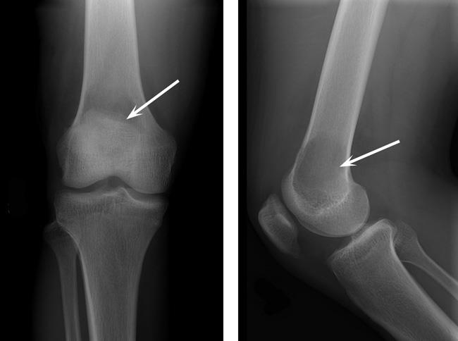
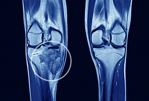
Xray images of bone cancer. Your doctor will examine other parts of your body to rule out cancers that can spread to bone. While a doctor may be able to see a tumor the x-ray will only tell the doctor if its there not if its malignant cancer or benign not cancer. CT MRI and PET scans can also tell if your cancer spread.
There is no special preparation for an x-ray. An X-ray is a procedure where radiation is used to produce images of the inside of the body. If an X-ray suggests you may have bone cancer.
The cancerous tumours yellow are in the metacarpal palm and phalanges finger and thumb bones. In fact noncancerous bone tumors are much more common than cancerous ones. Ewing sarcoma also is more likely to be in kids and young adults.
Swelling of the bone. Rottweilers In particular seem to be over-represented as a breed predisposed to bone cancer. The scans show the lymph nodes and distant organs where there might be cancer spread.
Take your medicines as normal. Bone cancer is rare making up less than 1 percent of all cancers. The four most common types of.
These are secondary tumours and have spread to the hand from a primary bone cancer in the leg. An x-ray is often the first test a doctor will order. 116649076 - Thoughtful young girl suffering from bone cancer wearing blue.
However malignant cells can also spread to the jaw from other cancers in the neck and head termed as secondary jaw cancer. Osteosarcoma is an aggressive cancer that spreads quickly to other parts of the body long before it is detected. For instance a lower gastrointestinal GI series often called a barium enema exam takes x-ray pictures after the bowel is filled with barium sulfate.
In most cases your doctor will order an x-ray to help diagnose a bone tumor. Plain X-rays will show bone mets only if they are more advanced go here. While x-ray images are among the clearest most detailed views of bone they provide little information about muscles tendons or joints.
CT scans are helpful in staging cancer. 147065056 - x-ray images chest or lung of patient for medical diagnose. Different types of tumors may look different.
Find bone cancer stock images in HD and millions of other royalty-free stock photos illustrations and vectors in the Shutterstock collection. Computed tomography CT scans. This is also known as primary bone cancerPrimary bone tumors are tumors that arise in the bone tissue itself and they may be benign or malignant bone cancerBenign non-cancerous tumors in the bones are more common than bone cancers.
Bone cancer can be found in cats as well but it is rare. When cancer is detected in bones it either originated in. Add to Likebox 149557327.
Special types of x-ray tests called contrast studies use iodine-based dyes or contrast materials like barium along with the x-rays to make the organs show up on the x-ray and get better pictures. During a bone scan a small amount of radioactive material is injected into your veins. Large and giant breed dogs have the greatest incidence of bone cancer in their limbs.
Bone cancer can begin in any bone in the body but it most commonly affects the pelvis or the long bones in the arms and legs. X-ray of lung showing chest cancer - bone cancer stock pictures royalty-free photos images an asian malay male patient prepared for hospital ct scan lying on the bed while a malay female nurse putting on a blanket for him - bone cancer stock pictures royalty-free photos images. A bone scan often shows metastases earlier than an X-ray and can check your whole body at once.
They help show if the bone cancer has spread to your lungs liver or other organs. Add to Likebox 113820794 - Bright photo of happy pretty girl suffering from kidney cancer. You can eat and drink normally beforehand.
Many bone cancers will show up on an x-ray. Images and X-rays of Bone Metastases normal anatomy. Swelling in the soft tissues surrounding the bone.
The tumor when it is located in this position can cause problems when moving severe random or constant pain and damage to the pelvic bone if the condition. The spread of a malignant cancer from one part of the body to another is known as metastasis. Pelvic bone cancer is a fairly rare medical condition that causes severe pain within the pelvisThis affliction begins with a small tumor forming upon the pelvis and as the condition becomes worse the tumors continue to grow in size.
X-rays provide images of dense structures such as bone. A chest x-ray is often done to see if bone cancer has spread to the lungs. Osteosarcoma the most common bone cancer usually happens to people ages 10 to 30 and most often starts in the arms legs or pelvis.
When you arrive the radiographer might ask you. A bone scan or CT are more sensitive than plain Xrays see right hip met and see rib metsAdvanced spine mets can lead to. What happens Before your x-ray.
A break in the bone fracture Preparing for your x-ray. Thousands of new high-quality pictures added every day. An MRI may be more useful in identifying bone and joint injuries eg meniscal and ligament tears in the knee rotator cuff and labrum tears in the shoulder and in imaging of the spine because both the bones and the spinal cord can be evaluated.
Bone cancer in the bones of the hand coloured X- ray. Other types of malignant cells that arise in the jaw bone are Ewings sarcomas or giant cell tumorsCancer that arises from the jaw bone is termed primary jaw cancer. Bone cancer is a malignant tumor that arises from the cells that make up the bones of the body.
