What New Coronavirus Looks Like Under The Microscope The images were made using scanning and transmission electron microscopes at the National Institute of Allergy and Infectious Diseases. Backscattered electron detector BSED.

It is a huge magnification of the common dust of industrial outdoor environments.
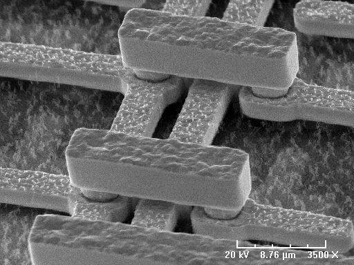
Scanning electron microscope images. Electromagnetic lenses Condenser lens Objective lens. The high-resolution three-dimensional images produced by SEMs provide topographical morphological and compositional information makes them invaluable in a variety of science and industry applications. The electron beam is scanned in a raster scan pattern and the position of.
Electronic Gun For the source of the electron. What youre seeing above is a scanning electron microscope image in false colour showing the COVID-19 virus from a patient in the US. As the intensity of the generated secondary electrons varies depending on the angle of the incident electrons onto the specimen surface subtle variations in the roughness of the surface can be expressed according to the signal intensity.
The electron source and electromagnetic lenses that generate and focus the beam are similar to those described for the. See more ideas about microscopic photography scanning electron microscope electron microscope. Feb 1 2017 - Explore Sara Kuntzs board Scanning Electron Microscope Images on Pinterest.
The scanning electron microscope SEM uses a focused beam of high-energy electrons to generate a variety of signals at the surface of solid specimens. - scanning electron microscope stock pictures royalty-free photos images. Colorful image for tops t-shirts skirts scarves leggings socks.
Thousands of new high-quality pictures added every day. Component or instrument used in scanning electron microscope. See more ideas about scanning electron microscope electron microscope images electron microscope.
Common industrial dust Technically this image is not a photograph since it was not originated by light photo but by an electron beam. Scanning electron microscopy SEM is a technique that provides high-resolution electronic images of the surface of different materials by scanning with an electron beam. The electrons in the beam interact with the sample producing various signals that can be used to obtain information about the surface topography and composition.
X-rays or light detector. A Scanning Electron Microscope SEM is a powerful magnification tool that utilizes focused beams of electrons to obtain information. Colourised sem image of atropa pollen showing the hard coat which protects the sperm cells during the movement from stamens of producing flower to the pistil of the receiving flower.
The signals that derive from electron-sample interactions reveal information about the sample including external morphology texture chemical composition and crystalline structure and orientation of materials making up the sample. Apr 22 2017 - Scanning Electron Microscope Images - Unbelievable how life really is up close. The Scanning Electron Microscope SEM normally detects secondary electrons to form an image for observation.
The viral particles are coloured yellow as it emerges from the surface of a cell which is coloured blue and pink. A scanning electron microscope SEM scans a focused electron beam over a surface to create an image. NIAID-RML The image above was captured with a transmission electron microscope.
25 Amazing Electron Microscope Images Writen by Bogdan Comments Off on 25 Amazing Electron Microscope Images All the common objects are kinda boring when you look at them but the situation changes when an awesome Electron Microscope comes in the scene. A scanning electron microscope SEM is a type of electron microscope that produces images of a sample by scanning the surface with a focused beam of electronsThe electrons interact with atoms in the sample producing various signals that contain information about the surface topography and composition of the sample. Secondary electron detector SED.
The image is modified and credit goes to Wikimedia. Atropa is a genus of the nightshade family. The image was captured by an Hitachi ultra-high-resolution Analytical FE Scanning Electron Microscope SU-70.
This scanning electron microscope image shows SARS-CoV-2 yellow the virus that causes COVID-19 emerging from the surface of cells bluepink cultured in the lab. Find scanning electron microscope stock images in HD and millions of other royalty-free stock photos illustrations and vectors in the Shutterstock collection. Scanning electron microscope SEM type of electron microscope designed for directly studying the surfaces of solid objects that utilizes a beam of focused electrons of relatively low energy as an electron probe that is scanned in a regular manner over the specimen.
In addition the contrast of the images provides information about the composition of the surface sample as its different elements emit different amounts of characteristic electrons. Scanning electron microscopy SEM in particular has given us some striking images over the years to tantalize our visual senses.
One advantage of ultrasound technology is that it allows substantial freedom in obtaining breast images from any orientation. Fatty breast tissue appears grey or black on images while dense tissues such as glands are white.
Pictures show breast structure and tumors.
Images of breast cancer. 15029 breast cancer awareness stock photos are available royalty-free. A woman with cancer is next to her daughter. 20619 breast cancer stock photos are available royalty-free.
This video aired in 2007 and yet when I shared it with my close friends none of them had ever heard of it. The above ultrasound images show a typical proven case of cancer of the left breast. However you should be careful to notice it because people often get confused with a breast infection with this disease.
- women breast cancer stock pictures royalty-free photos images. Confident woman standing in city for breast cancer awareness. Generally speaking the denser the tissue the whiter it appears.
This may include normal tissue and glands as well as areas of benign breast changes eg fibroadenomas and disease breast cancerFat and other less-dense tissue renders gray on a mammogram image. Download Breast cancer stock photos. The symptoms of the Inflammatory Breast Cancer are highly visible and different from the other breast cancers.
Reset All Filters October Breast Cancer Awareness month adult Woman hand holding Pink Ribbon on pink background for supporting people living and. Inflammatory breast cancer is an infrequent aggressive type of breast cancer that spreads rapidly. Doctor holds pink ribbon international breast cancer day.
Its not pretty but its critical information. Breast cancer starts when normal cells transform into cancerous cells and these breast cells begin to grow out of control. Power to fight breast cancer The power to fight breast cancer women wearing pink ribbons for breast cancer campaign on white background breast cancer stock pictures royalty-free photos images An Asian woman in her 60s embraces her mid-30s daughter who is battling cancer An ethnic woman wearing a headscarf and fighting cancer sits on the couch with her mother.
After some time breast cancerous cells form a tumor breast lump that can be felt and identified by palpation and could be seen on mammogram. Affordable and search from millions of royalty free images photos and vectors. Its the most common cancer in women although it can also develop in men.
The exact cause of breast. Inflammatory breast cancer pictures show a red andor swollen breast that appears inflamed. However some breast cancer tumors.
Pictures Of Inflammatory Breast Cancer The IBC Network Admin 2021-01-08T175007-0600 There are images below on this page that contain graphic medical photos. Breast cancer cells can invade into surrounding tissues or spread to other areas. If a doctor suspects an abnormal growth in the breast after a clinical breast examination or screening mammography evaluation of breast cancer ultrasound images will help confirm the diagnosis.
Cells also blocks the lymph vessels located in the skin of the breast. A mammogram image has a black background and shows the breast in variations of gray and white. This video shows photos of inflammatory breast cancer.
The Inflammatory Breast Cancer turns the affected breast red and painful. We are sharing these photos for educational purposes. In addition the mass shows multiple echogenic areas along the rim a clear sign of malignancy in breast carcinoma.
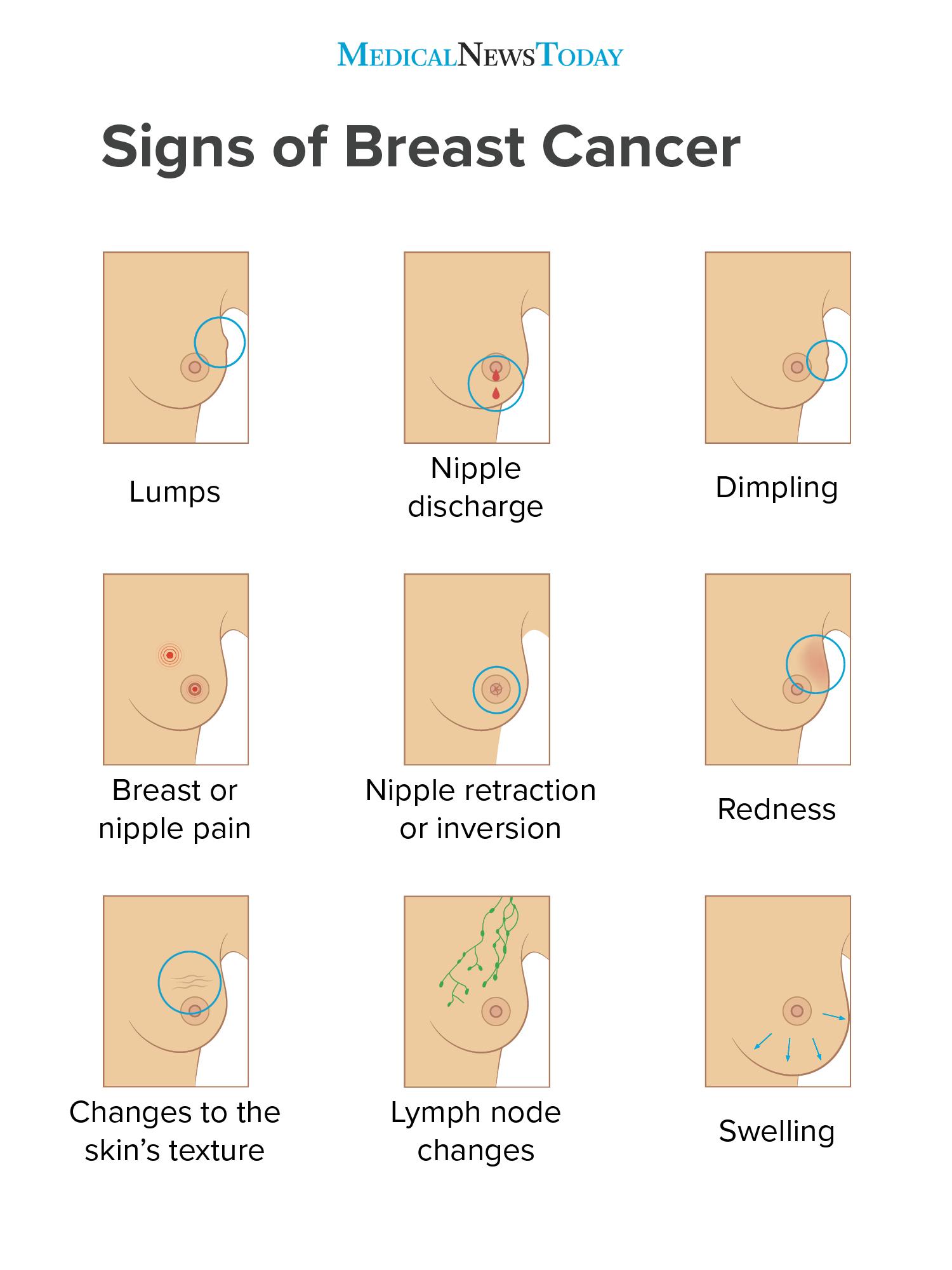
The ACS report that these lumps are usually hard irregular in shape and painless. Most cases are invasive ductal carcinomas which develop in the cells lining the milk ducts and spread throughout the breast. So few people have heard of this type of cancer.
The tumor is seen as a well defined hypoechoic mass with microlobulation or fine irregularities of the margins. A girl is hugging a woman happy - women breast cancer stock pictures royalty-free photos images. Learn about the breast cancer experience from symptoms and tests to treatments recovery and prevention.
Check Inflammatory Breast Cancer Pictures images to examine itchy rash bruises red spots discoloration or pain in breasts with early signs symptoms. October Breast Cancer Awareness month adult Woman hand holding Pink Ribbon on pink background for supporting people living and. A new mass or lump in breast tissue is the most common sign of breast cancer.
A mammogram can help a doctor to diagnose breast cancer or monitor how it responds to treatment. Breast cancer is the uncontrollable growth of malignant cells in the breasts.
Be ready for an x-ray. A bone scan can give more detailed information about the inside of your bones than an X-ray.
Picture of a bone scan showing hot spots black areas which are sites of bone cancer see the close up of bone scan and here and here.
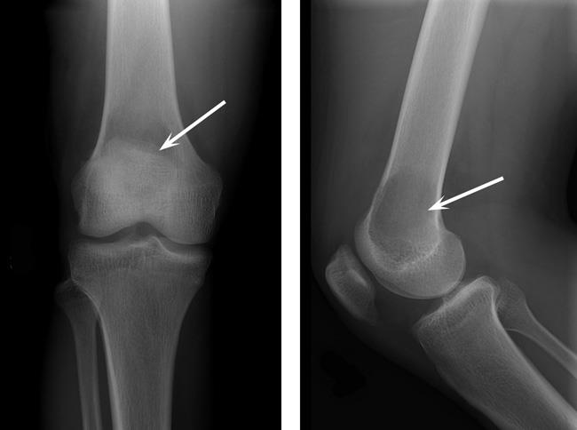
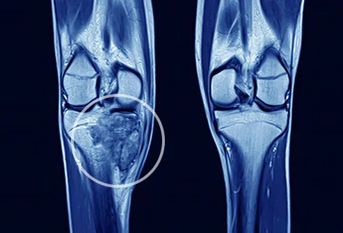
Xray images of bone cancer. Your doctor will examine other parts of your body to rule out cancers that can spread to bone. While a doctor may be able to see a tumor the x-ray will only tell the doctor if its there not if its malignant cancer or benign not cancer. CT MRI and PET scans can also tell if your cancer spread.
There is no special preparation for an x-ray. An X-ray is a procedure where radiation is used to produce images of the inside of the body. If an X-ray suggests you may have bone cancer.
The cancerous tumours yellow are in the metacarpal palm and phalanges finger and thumb bones. In fact noncancerous bone tumors are much more common than cancerous ones. Ewing sarcoma also is more likely to be in kids and young adults.
Swelling of the bone. Rottweilers In particular seem to be over-represented as a breed predisposed to bone cancer. The scans show the lymph nodes and distant organs where there might be cancer spread.
Take your medicines as normal. Bone cancer is rare making up less than 1 percent of all cancers. The four most common types of.
These are secondary tumours and have spread to the hand from a primary bone cancer in the leg. An x-ray is often the first test a doctor will order. 116649076 - Thoughtful young girl suffering from bone cancer wearing blue.
However malignant cells can also spread to the jaw from other cancers in the neck and head termed as secondary jaw cancer. Osteosarcoma is an aggressive cancer that spreads quickly to other parts of the body long before it is detected. For instance a lower gastrointestinal GI series often called a barium enema exam takes x-ray pictures after the bowel is filled with barium sulfate.
In most cases your doctor will order an x-ray to help diagnose a bone tumor. Plain X-rays will show bone mets only if they are more advanced go here. While x-ray images are among the clearest most detailed views of bone they provide little information about muscles tendons or joints.
CT scans are helpful in staging cancer. 147065056 - x-ray images chest or lung of patient for medical diagnose. Different types of tumors may look different.
Find bone cancer stock images in HD and millions of other royalty-free stock photos illustrations and vectors in the Shutterstock collection. Computed tomography CT scans. This is also known as primary bone cancerPrimary bone tumors are tumors that arise in the bone tissue itself and they may be benign or malignant bone cancerBenign non-cancerous tumors in the bones are more common than bone cancers.
Bone cancer can be found in cats as well but it is rare. When cancer is detected in bones it either originated in. Add to Likebox 149557327.
Special types of x-ray tests called contrast studies use iodine-based dyes or contrast materials like barium along with the x-rays to make the organs show up on the x-ray and get better pictures. During a bone scan a small amount of radioactive material is injected into your veins. Large and giant breed dogs have the greatest incidence of bone cancer in their limbs.
Bone cancer can begin in any bone in the body but it most commonly affects the pelvis or the long bones in the arms and legs. X-ray of lung showing chest cancer - bone cancer stock pictures royalty-free photos images an asian malay male patient prepared for hospital ct scan lying on the bed while a malay female nurse putting on a blanket for him - bone cancer stock pictures royalty-free photos images. A bone scan often shows metastases earlier than an X-ray and can check your whole body at once.
They help show if the bone cancer has spread to your lungs liver or other organs. Add to Likebox 113820794 - Bright photo of happy pretty girl suffering from kidney cancer. You can eat and drink normally beforehand.
Many bone cancers will show up on an x-ray. Images and X-rays of Bone Metastases normal anatomy. Swelling in the soft tissues surrounding the bone.
The tumor when it is located in this position can cause problems when moving severe random or constant pain and damage to the pelvic bone if the condition. The spread of a malignant cancer from one part of the body to another is known as metastasis. Pelvic bone cancer is a fairly rare medical condition that causes severe pain within the pelvisThis affliction begins with a small tumor forming upon the pelvis and as the condition becomes worse the tumors continue to grow in size.
X-rays provide images of dense structures such as bone. A chest x-ray is often done to see if bone cancer has spread to the lungs. Osteosarcoma the most common bone cancer usually happens to people ages 10 to 30 and most often starts in the arms legs or pelvis.
When you arrive the radiographer might ask you. A bone scan or CT are more sensitive than plain Xrays see right hip met and see rib metsAdvanced spine mets can lead to. What happens Before your x-ray.
A break in the bone fracture Preparing for your x-ray. Thousands of new high-quality pictures added every day. An MRI may be more useful in identifying bone and joint injuries eg meniscal and ligament tears in the knee rotator cuff and labrum tears in the shoulder and in imaging of the spine because both the bones and the spinal cord can be evaluated.
Bone cancer in the bones of the hand coloured X- ray. Other types of malignant cells that arise in the jaw bone are Ewings sarcomas or giant cell tumorsCancer that arises from the jaw bone is termed primary jaw cancer. Bone cancer is a malignant tumor that arises from the cells that make up the bones of the body.
The type of cells involved in your tongue cancer helps determine your prognosis and treatment. The photos below give you an idea of what tongue cancers can look like but remember that they might appear differently from this.
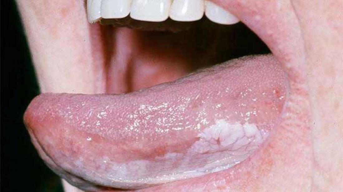 Tongue Cancer Symptoms Pictures And Outlook
Cancer of the tongue is a common yet severe subtype of head and neck cancer.

Images of tongue cancer. Pics Images Pictures and Photos of Mouth Cancer. Tongue Cancer Survival Rate. Oral cancer affects thousands of people every year.
Oral cancer affects the lips gums tongue roof of the mouth insides of the cheeks or the soft floor of the mouth under the tongue. Tongue Cancer Pictures Photos and Images Early signs stages under tongue hpv symptoms treatment. Prevention within Early Tongue Cancer Pictures Article Related to Early Tongue Cancer Pictures.
Pictures of mouth cancer These photos give you an idea of what possible mouth cancers can look like but remember that they might appear differently to this. Here are some photos of those diseases. If you can get past that theres always the threat of cancer and other oral diseases.
Tobacco use whether smoking or chewing can also lead to cancer of the lips as well as other mouth cancers. Check out some pictures to know what to look out for and find out about the signs and symptoms of mouth cancer in the lips teeth and gums. Tongue cancer is a type of cancer that starts in the cells of the tongue and can cause lesions or tumors on your tongue.
Symptoms Pictures Treatment Causes. Some contain a brief patient history which may add insight to the actual diagnosis of the disease. Its important to be aware of the symptoms of mouth cancer so you can contact your GP or dentist if you notice anything abnormal.
Mouth cancer is a type of head and neck cancer and it often comes under the category of oral and oropharyngeal cancerOral cancer accounts for roughly 3 of all cancer diagnoses in the United. Around 8300 people are diagnosed with mouth cancer each year in the UK which is about 1 in every 50 cancers diagnosed. Its a type of head and neck cancer.
Lip cancer lesions can develop anywhere on your lips but they are most likely to occur on your lower lip. See oral cancer stock video clips. Only 1 in 8 125 happen in people younger than 50.
Diseases Conditions Rashes Syndromes Skin Conditions. The survival rate of tongue cancer is 50 percent. Tongue cancer light micrograph - mouth cancer stock pictures royalty-free photos images we have to protect the life from coronavirus - mouth cancer stock pictures royalty-free photos images In the 1970s UNICEF initiated the boring of deep wells in villages across the Ganges Delta in a well intentioned mission to eradicate water born.
1 BSIP Medical Images. Advanced stage of Cancer with Gum Affectation. Click on each photo for a.
The most noticeable signs of. Tongue cancer is a type of mouth cancer or oral cancer that usually develops in the squamous cells on the surface of the tongueIt can cause tumors or lesions. Mouth Oral Cancer Stages.
Smokeless tobacco dip snuff and chew are dangerous. Find tongue cancer stock images in HD and millions of other royalty-free stock photos illustrations and vectors in the Shutterstock collection. More than 2 in 3 cases of mouth cancer develop in adults over the age of 55.
Mouth cancer is the 6th most common cancer in the world but its much less common in the UK. Hopefully this isnt you. Storey of Someone Tongue Cancer A Killer Developing In My Mouth early tongue cancer pictures I cant sleepMy mind saunters with the things that are to comeI was diagnosed a pair weeks ago with stage 3 tongue cancer.
Home Dental Oral Cancer Images This collection of photos contains both cancer and non-cancerous diseases of the oral environment which may be mistaken for malignancies. If there is a localized cancer there is high rate for a five-year survival. Thousands of new high-quality pictures added every day.
2319 oral cancer stock photos vectors and illustrations are available royalty-free. Getting too much sun is a risk factor for oral cancer especially lip cancerMaking sure you have sun-protectant lip balm or lipstick is one way to cut your risk. Now the cancer cells multiply erratically thus growing into a tumor affecting one particular area where it is attached.
However if the cancer has metastasized there is a lower survival rate for persons with tongue cancer. Mouth cancer mucositis oral cancer awareness dental cancer head and neck tumor international cancer oral mucosa infographic teeth grind teeth brushing gums. Not only do they cause bad breath sore throats they are incredibly disgusting.
Tongue cancer is a form of cancer that begins in the cells of the tongue. Several types of cancer can affect the tongue but tongue cancer most often begins in the thin flat squamous cells that line the surface of the tongue. Contact your GP or dentist if you notice anything abnormal.
It is the initial stage of mouth cancer where small cancer or tumor is present in one area of the mouth. Find out about mouth cancer staging. Tongue cancer that starts in the front two thirds of your tongue oral tongue is staged as a mouth cancer.
Try these curated collections.

